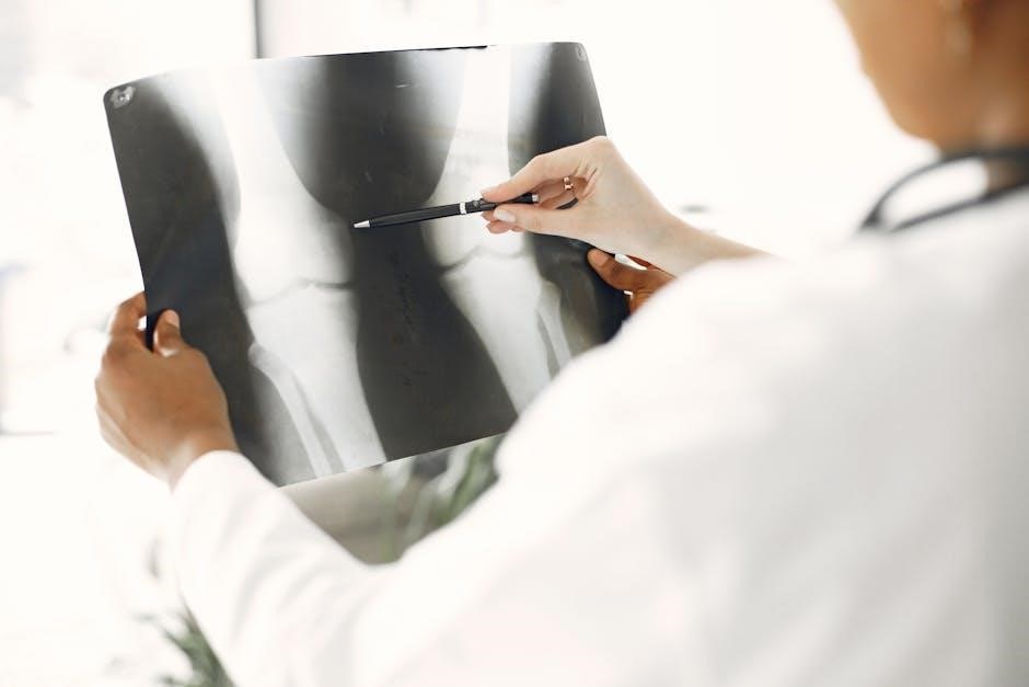
Bone fractures, detailed in numerous PDF resources, represent disruptions to bone continuity; understanding these breaks—transverse, oblique, spiral, and comminuted—is crucial.
Fracture classification, often found within medical PDF guides, aids diagnosis and treatment planning, encompassing closed, open, stable, and unstable fracture types.
What is a Bone Fracture?
A bone fracture signifies a break in the continuity of the bone, ranging from a hairline crack to a complete shatter. Numerous PDF documents detail these injuries, categorizing them based on various factors. These resources illustrate that fractures occur when physical force exceeds the bone’s structural strength.
PDF guides commonly outline fracture patterns like transverse (straight across), oblique (angled), spiral (twisting), and comminuted (multiple fragments). Understanding these types, as presented in medical literature available in PDF format, is vital for accurate diagnosis. Further, PDFs explain distinctions between closed (skin intact) and open (skin penetrated) fractures, impacting treatment protocols; The severity and treatment depend heavily on the fracture’s specific characteristics, thoroughly documented in accessible PDF materials.
Importance of Fracture Classification
Accurate fracture classification, extensively detailed in medical PDFs, is paramount for effective patient care. These PDF resources demonstrate how categorizing fractures – by pattern (transverse, spiral, etc.), skin penetration (open/closed), and stability – directly influences treatment decisions.
PDF guides emphasize that proper classification guides appropriate immobilization techniques, surgical interventions, and rehabilitation protocols. Understanding the type of fracture—whether a simple transverse break or a complex comminuted fracture—dictates the urgency and complexity of care. Accessing comprehensive PDF materials allows healthcare professionals to consistently apply standardized classification systems, improving communication and ensuring optimal outcomes. Detailed PDFs also aid in predicting potential complications based on fracture characteristics.
Types of Bone Fractures Based on Pattern
PDF resources categorize fractures by pattern: transverse, oblique, spiral, segmental, and comminuted, detailing each type’s characteristics and implications for treatment.
Transverse Fractures
Transverse fractures, thoroughly documented in orthopedic PDF guides, present as a straight break across the bone’s axis, perpendicular to its length. These fractures typically result from direct force impacting the bone, such as a fall or a direct blow. PDF resources emphasize that diagnosis often involves radiographic imaging to confirm the clean, horizontal fracture line.
Treatment protocols, detailed in specialized PDFs, vary based on fracture location and severity, ranging from immobilization with casts to surgical intervention involving internal fixation. Understanding the stability of a transverse fracture—whether it’s stable or unstable—is crucial, as outlined in medical PDFs, influencing the chosen treatment pathway. Proper alignment and stabilization are key to successful healing, as illustrated in numerous clinical PDF case studies.
Oblique Fractures
Oblique fractures, extensively illustrated in orthopedic PDF materials, are characterized by a break line angled across the bone, not perpendicular like transverse fractures. These often occur due to twisting forces or angled impacts, as detailed in trauma PDF reports. Diagnostic PDFs highlight the importance of multiple radiographic views to accurately assess the fracture’s angle and displacement.
Treatment strategies, outlined in surgical PDF guides, frequently involve reduction – realigning the bone fragments – followed by immobilization or surgical fixation. The instability of an oblique fracture, a key factor discussed in clinical PDFs, dictates the need for more robust stabilization methods. PDF resources emphasize that proper reduction and fixation are vital to restore optimal function and prevent malunion, with post-operative rehabilitation protocols also detailed.
Spiral Fractures
Spiral fractures, thoroughly documented in orthopedic PDFs, result from a twisting injury applied to a bone. These fractures exhibit a helical pattern around the bone’s axis, often seen in sports injuries or abuse cases, as detailed in forensic PDF reports. Radiographic assessments, explained in diagnostic imaging PDFs, are crucial for identifying the fracture’s complete extent and rotational displacement.
Treatment approaches, outlined in surgical PDF guides, typically involve reduction and stabilization, frequently requiring internal fixation with plates or rods. The inherent instability of spiral fractures, emphasized in clinical PDFs, often necessitates surgical intervention. PDF resources highlight the importance of addressing any associated soft tissue injuries alongside the fracture repair, ensuring optimal healing and functional recovery through rehabilitation protocols.
Comminuted Fractures
Comminuted fractures, extensively illustrated in orthopedic trauma PDFs, are characterized by the bone being broken into three or more fragments. These complex injuries, detailed in surgical technique PDFs, often result from high-energy trauma like vehicle accidents or falls. Diagnostic imaging PDFs emphasize the necessity of comprehensive radiographic evaluation, including CT scans, to fully assess fragment size and displacement.
Treatment, as described in advanced fracture care PDFs, usually requires open reduction and internal fixation (ORIF) utilizing plates, screws, or external fixators. Reconstruction PDFs highlight the challenges of restoring anatomical alignment and achieving stable fixation. Rehabilitation protocols, found in post-operative care PDFs, are crucial for regaining function, often requiring prolonged therapy to address stiffness and weakness.
Segmental Fractures
Segmental fractures, thoroughly documented in orthopedic PDF resources, involve a bone broken in two places, with a floating segment of bone between the fracture lines. These fractures, often visualized in radiographic imaging PDFs, present unique challenges due to the instability of the intermediate bone segment. Treatment planning PDFs emphasize the need for stabilization of both fracture sites to prevent malunion or nonunion.
Surgical intervention, detailed in operative technique PDFs, typically involves ORIF with bridging plates or intramedullary nails to span the fracture gap. Rehabilitation guidelines, found in post-operative care PDFs, focus on restoring range of motion and strength, often requiring a prolonged and carefully monitored recovery period. Case study PDFs illustrate the complexities of managing these fractures.

Types of Bone Fractures Based on Skin Penetration

PDF guides categorize fractures as either closed (skin intact) or open (skin penetrated), impacting infection risk and treatment protocols, as detailed in medical PDFs.
Closed (Simple) Fractures
Closed fractures, extensively documented in orthopedic PDF resources, are breaks where the bone hasn’t pierced the skin. These fractures are characterized by pain, swelling, and deformity, but without an open wound communicating with the fracture site. Medical PDFs emphasize that diagnosis typically involves X-rays to confirm the break and assess its severity.
Treatment, as outlined in various PDF guides, usually involves immobilization with a cast or splint to allow the bone to heal naturally. While generally less prone to infection than open fractures, proper alignment and stabilization are crucial for optimal healing, as detailed in rehabilitation PDFs. PDFs also highlight the importance of pain management and monitoring for complications.
Further information regarding specific closed fracture types and treatment protocols can be found within comprehensive orthopedic trauma PDF manuals.
Open (Compound) Fractures
Open fractures, thoroughly described in emergency medicine PDFs, are characterized by a broken bone protruding through the skin, or a wound that connects to the fracture site. These injuries carry a significantly higher risk of infection, as detailed in surgical PDF guides, due to potential bacterial contamination. Immediate medical attention is critical, as emphasized in trauma care PDFs.
Treatment, as outlined in orthopedic PDF resources, involves surgical intervention to clean the wound, stabilize the fracture—often with internal fixation—and prevent infection. Antibiotics are administered, and tetanus prophylaxis is essential. Rehabilitation PDFs detail the lengthy recovery process, often involving physical therapy.
Detailed classifications of open fracture severity (Gustilo-Anderson) are readily available in specialized orthopedic PDFs, guiding treatment decisions and predicting outcomes.

Types of Bone Fractures Based on Stability
PDF resources categorize fractures as stable—aligned and minimally displaced—or unstable, requiring intervention; stability impacts treatment protocols and healing potential.
Stable Fractures
Stable fractures, extensively documented in orthopedic PDF guides, demonstrate minimal displacement of bone fragments, maintaining reasonable alignment. These fractures generally occur when the bone is broken, but the pieces remain in acceptable position.
PDF resources emphasize that stable fractures often result from lower-energy impacts and typically don’t disrupt surrounding soft tissues significantly. Treatment frequently involves immobilization—casts or splints—allowing the bone to heal naturally without surgical intervention.
However, PDF-based clinical guidelines stress the importance of radiographic evaluation to confirm stability and rule out subtle displacements. Monitoring for any signs of instability during the healing process, as detailed in specialized PDF reports, is also crucial for optimal outcomes.
Unstable Fractures

Unstable fractures, thoroughly illustrated in orthopedic PDF manuals, exhibit significant displacement or fragmentation of bone, compromising structural integrity. These fractures frequently require intervention to restore proper alignment and stability, as detailed in surgical PDF guides.
PDF resources highlight that unstable fractures often result from high-energy trauma, frequently involving comminution or extension into joint spaces. They pose a higher risk of non-union or malunion if not appropriately managed.
Treatment typically involves surgical fixation—plates, screws, or rods—to maintain reduction during healing, a process extensively covered in PDF-based surgical protocols. Post-operative immobilization and rehabilitation, as outlined in physiotherapy PDFs, are essential for restoring function and preventing complications.
Specific Fracture Types & Mechanisms
PDF guides detail specific fractures—stress, avulsion, impacted, and compression—explaining their unique mechanisms and characteristics for accurate diagnosis and treatment planning.
Stress Fractures
Stress fractures, thoroughly documented in orthopedic PDF resources, are small cracks developing in bone due to repetitive stress, rather than a single traumatic event; These often occur in weight-bearing bones of the lower leg and foot, frequently seen in athletes.
PDF guides emphasize that stress fractures are initially difficult to detect on standard X-rays; bone scans or MRIs, as detailed in specialized PDFs, are often required for diagnosis. Treatment, outlined in rehabilitation PDFs, typically involves rest, activity modification, and gradual return to function.
Understanding the biomechanics contributing to stress fractures, as explained in sports medicine PDFs, is crucial for prevention, including proper training techniques and footwear. Ignoring early symptoms can lead to more significant fractures, as warned in comprehensive fracture PDFs.
Avulsion Fractures
Avulsion fractures, clearly illustrated in orthopedic PDF manuals, occur when a tendon or ligament pulls a small piece of bone away from the main bone mass. These are frequently seen around joints, particularly in adolescents whose bones are still developing, as detailed in pediatric fracture PDFs.
PDF resources highlight common locations for avulsion fractures, including the tibial tuberosity and the hip. Diagnosis, as described in radiology PDFs, often involves X-rays, though sometimes MRI is needed to assess soft tissue damage. Treatment, outlined in rehabilitation PDFs, typically involves immobilization and physical therapy.
Understanding the mechanism of injury – a forceful muscle contraction – is key, as explained in sports medicine PDFs. Preventing avulsion fractures involves proper warm-up and conditioning, as emphasized in preventative care PDF guides.
Impacted Fractures
Impacted fractures, thoroughly documented in orthopedic trauma PDFs, occur when one fragment of a broken bone is driven into another. These fractures, often seen in long bones, are characterized by a shortening of the bone length, as illustrated in surgical PDF guides.
PDF resources emphasize that impacted fractures frequently result from axial loading or high-energy trauma. Radiographic assessment, detailed in radiology PDFs, reveals the telescoping of bone fragments. Treatment, as described in post-operative care PDFs, often requires surgical intervention to restore bone length and alignment.
Internal fixation, using plates and screws, is commonly employed, as shown in surgical technique PDFs. Understanding the stability of the impactation is crucial for treatment planning, per fracture management PDFs.
Compression Fractures
Compression fractures, extensively covered in spine-focused PDFs, typically affect vertebral bodies, often due to osteoporosis or significant trauma. These fractures, detailed in orthopedic PDF guides, involve the collapse of bone, reducing vertebral height, as visualized in imaging PDFs.
PDF resources highlight that compression fractures commonly present with acute back pain. Diagnosis relies on X-rays and MRI, explained in radiology PDFs, to assess the degree of vertebral collapse. Treatment, outlined in rehabilitation PDFs, ranges from conservative management—bracing and pain control—to surgical stabilization.
Kyphoplasty and vertebroplasty, detailed in interventional radiology PDFs, are minimally invasive procedures used to restore vertebral height and reduce pain, as shown in procedural PDFs.

Resources for Further Information (PDF Focus)
Numerous medical websites and institutions offer comprehensive PDF guides detailing types of bone fractures, aiding identification and understanding of treatment options.
Finding Reliable Fracture Information in PDF Format
Locating trustworthy information regarding types of bone fractures in PDF format requires careful source evaluation. Prioritize documents originating from reputable medical institutions, such as hospital websites (Mayo Clinic, Johns Hopkins), professional organizations (American Academy of Orthopaedic Surgeons ⸺ AAOS), and government health agencies (NIH, CDC).
Utilize advanced search operators (e.g., “bone fracture types” filetype:pdf) within search engines to refine results. Beware of websites with unclear authorship or commercial bias. Look for PDFs that are regularly updated and cite credible sources. Many university medical libraries also offer publicly accessible PDF resources on orthopedic injuries.
Always cross-reference information from multiple sources to ensure accuracy and a comprehensive understanding of fracture classifications – transverse, oblique, spiral, comminuted, and others – as detailed in reliable PDF guides.
Utilizing PDF Resources for Fracture Identification
PDF resources detailing types of bone fractures are invaluable for visual learning. Focus on diagrams and radiographic images illustrating fracture patterns – transverse, oblique, spiral, comminuted, and stress fractures. Pay attention to descriptions of fracture lines, displacement, and angulation.

Many PDF guides include case studies demonstrating fracture identification in clinical scenarios. Utilize these to practice recognizing different fracture types. Compare and contrast closed versus open fractures, and stable versus unstable configurations. Remember that accurate identification often requires professional medical expertise.
When reviewing PDFs, note the specific anatomical location of the fracture, as this influences treatment approaches. Supplement PDF study with interactive online resources and consult with healthcare professionals for definitive diagnoses.